Groundbreaking Vision Diagnostics at Pacific Vision Institute
Pacific Vision Institute is the first facility in Northern California and the only clinic in San Francisco to offer this advanced and accurate diagnostic scan in screening vision correction candidates. Drs. Faktorovich and the doctors at Pacific Vision Institute have presented and published extensively on the use of the corneal OCT scan to enhance accuracy in planning refractive surgery. This scan will be performed as part of your initial consultation.
How the Widefield OCT is Used
The OCT is a diagnostic tool for the advanced analysis of the anterior segment and corneal pachymetry (measuring the thickness of the cornea) but can have many other applications as well. The integrity of the cornea and the ocular surface is vital for functional vision. OCT technology can generate very high resolution, cross section and 3D images of the retina, optic disc and the anterior segment.
The scan is a simple procedure and takes just minutes. The results can be broken down to visualize individual retinal layers for increased diagnostic certainty and it gives our eye specialists the ability to see and track, over time, cross sections of the cornea, measure angles, and calculate thickness.
Cutting-Edge Evaluations for Superior Results in Vision Correction
This cutting-edge technology has given eye surgeons unprecedented access to the complex elements of the human eye and its microvasculature content and enables doctors to make more precise changes for better vision than has ever before been possible. With OCT technology, our team of eye specialists can evaluate a patient’s eye as often as needed to track the progression of an eye disease or condition to evaluate how well treatment is performing. Being able to see the progress of the eye in real time enormously increases the efficacy of the procedure selected and ultimately a successful outcome.
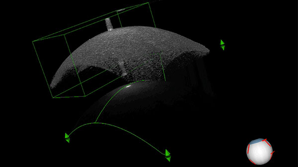
How Widefield OCT Works
OCT is an optical method of scanning that is based on light reflection and scattering from the elements within the cornea. When different reflectivity from the internal structures are measured, a cross-section image is produced. As a result, the reflecting structures’ locations can be accurately visualized with a high degree of resolution. OCT is non-contact, allowing structures such as the iris and lens to be seen in their natural state.
Advanced Imaging with Widefield OCT
The imaging is performed by gradually moving the point of focus forward in small increments to obtain a succession of coronal slices from the surface of the cornea to the back of the lens. When these coronal images are assembled into one view, it can be seen how changes in the eye are related to coronal elements in an easy-to-view 3D manner. The human eye is a complex organ loaded with elements of extremely fine tolerances.
Many of these elements were not able to be clearly observed previously. Imagine the difference in looking at the night sky with a pair of binoculars and then using a powerful telescope. This more comprehensive picture of pathologies in the eye and its progression takes a great deal of guesswork out of vision treatments.
Benefits of Widefield OCT
- Quick, safe, non-invasive and pain-free with no downtime and takes about ten minutes.
- Provides comprehensively detailed images of the cornea and retina and the sub-layers of the eye.
- Provides real-time imaging of the form and structure of tissue in the eye.
- Unprecedented image resolution helps educate patients on their condition and treatment.
- No preparation of patient required and no downtime.
- Essential for early diagnosis of glaucoma, macular degeneration and diabetic retinal disease.
Schedule a Consultation
Send us a message to schedule an in person consultation at our office in San Francisco.
Schedule